Computer Analysis
Computerized Varve Thickness Measurement
Measuring a varve record from a core involves measuring the thicknesses of summer and winter layers in successive couplets from bottom to top in the core. If this is done using a computer, a computer program must be able to load successive images of the varve core and allow the marking of boundaries that separate summer and winter layers. Simultaneously, the computer program must keep track of thicknesses in a spreadsheet and also allow for the posting of comments about individual varves as well as mark the beginning varve of each successive image with an image number. The advantage to doing this is that it creates an image and numerical record of varve measurements that can serve as an invaluable archive and source of information for further study. Below is an example of an analyzed image and data table produced by our program. Note that the data file lists varves from the bottom of the core at the top of the page and progresses down the page with each line representing successively younger varve measurements.
At Tufts University, varve measurement is performed with a script program written for UTHSCSA Image Tool 3.0. Image Tool is a free image processing and analysis program developed for medical applications in the Department of Dental Diagnostic Science at The University of Texas Health Science Center, San Antonio, Texas. The script program, Varve300.itm, takes advantage of utilities built into Image Tool. Varve300 loads Jpeg or Bitmap images and allows the operator to successively measure the varves on each image, while assembling a data table.
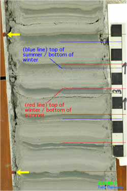
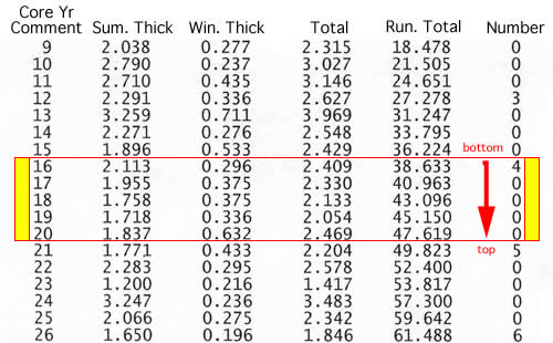
Left: red marks on left core liner (yellow arrows) = image range to be measured on this image. Right: data file result from core measurement. Note data file lists varves in reverse order as they are measured bottom to top. Core ALD6B, image 4 (ALD6B-4) from Aldrich Brook core site in Westmoreland, NH
Computerized Varve Gray-Scale Analysis
Measuring a varve record from a core involves measuring the thicknesses of summer and winter layers in successive couplets from bottom to top in the core. Digital gray-scale analysis can assist in making decisions about summer/winter layer boundaries as well as pulling out some of the details of bedding in the summer layer. The gray scale analysis is a less biased way of looking at varve images.
At Tufts University, varve gray-scale data is assembled with a script program written for UTHSCSA Image Tool 3.0. Image Tool is a free image processing and analysis program developed for medical applications in the Department of Dental Diagnostic Science at The University of Texas Health Science Center, San Antonio, Texas. The script program, Varveprofile100.itm, creates a gray-scale data file for pixels along a strip of specified pixel width and takes advantage of utilities built into Image Tool.
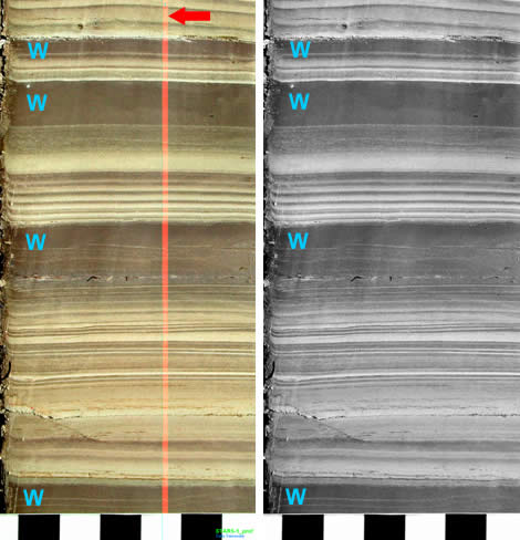
Left: High resolution color (RGB) image of dried varve core with image sampling profile line that is 20 pixels wide. (W = winter layers). Right: Gray scale version (total RGB values) of high resolution image.
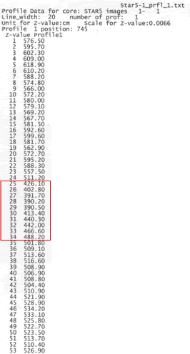
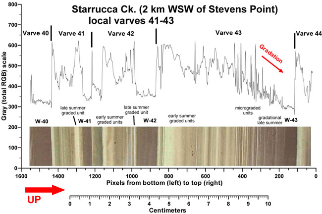
The top of the gray scale data file from the profile line (light vertical band) shown on the RGB image. Data are average total RGB values from 0 (black) to 768 (white) for each 20 pixel row on the profile line. The whole profile is 2008 pixels long with data from only the top 53 rows of the image shown here. The highlighted data is for the top-most dark lamination on the images (red arrow) that has lower RGB values than the surrounding sediment. Plotting the gray scale data gives a graphical representation of the thicknesses and gray scale contrasts of the layering in each varve. Below is an example of the gray scale profile of an image with several varves
Analysis Tools Downloads
Go to the link below to download the necessary software for a Windows operating system. Unfortunately we do not have a Mac version of the program. PLEASE read the instructions for setting up and using the varve measurement program before installing any of the programs!!
References
- Wilcox, C.D., Dove, S.B., McDavid, W.D., and Greer, D.B., 2002, UTHSCSA Image Tool Version 3.0: Freeware software available from the Department of Dental Diagnostic Science at the University of Texas Health Science Center at San Antonio: Download UTHSCSA Image Tool 3.0 (Or perform a web search for "Image Tool 3.0").


